


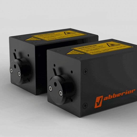
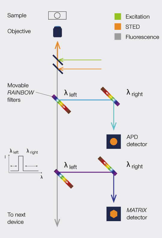
RAINBOW Detection
The abberior RAINBOW Detection pairs high transmission and quantum efficiency with optimal wavelength discrimination. There are no more compromises: you benefit from both maximum signal and full spectral flexibility! The superior detection efficiency of APDs compared to any other photon detector, including PMTs and hybrid detectors, guarantees that you collect as many precious fluorescence photons from your sample as possible.
full contrast, full flexibility
MATRIX Detector
Many eyes see more than one. The MATRIX detector drastically improves signal-to-background ratio, resolution, and dynamic range.
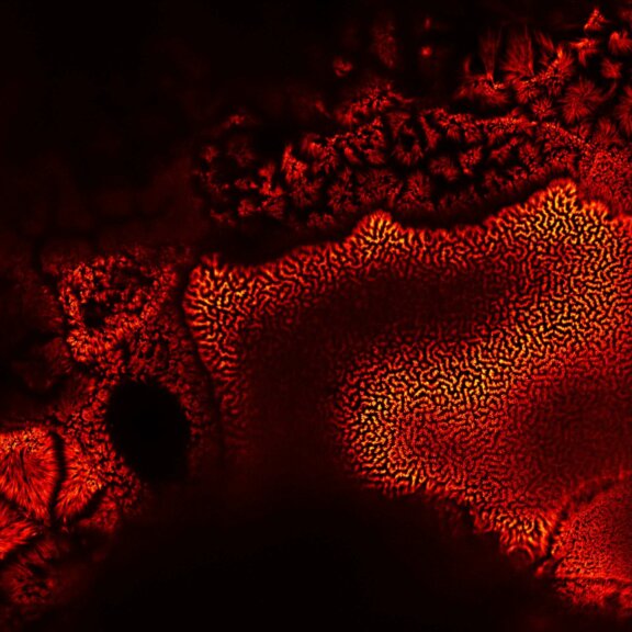
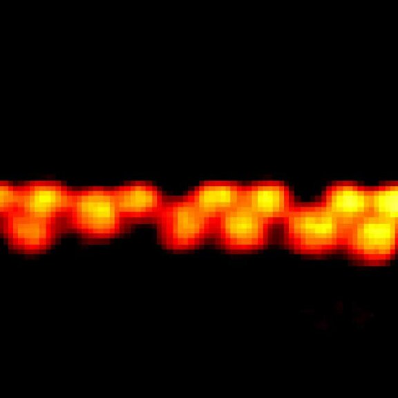
TIMEBOW Imaging
TIMEBOW lifetime imaging for stunning results at confocal and STED super-resolution.
FLEXPOSURE Illumination
Brings down the light dose on your sample and lables dramatically. Key ingredient for volume and live-cell superresolution.
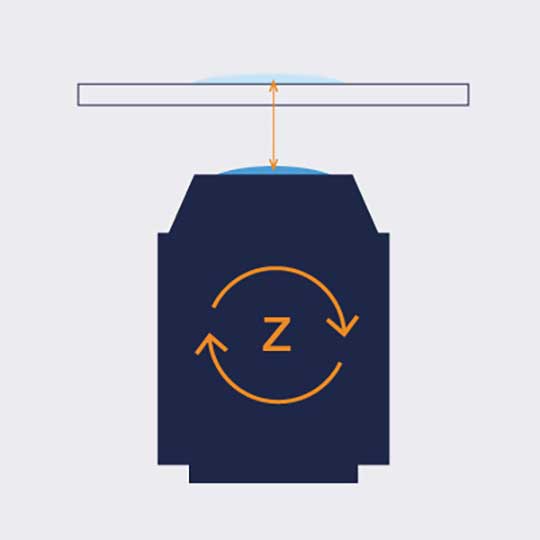
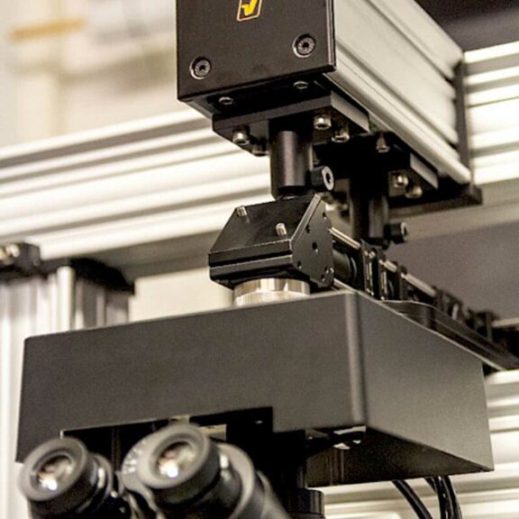
RAYSHAPE Mirror
Dynamic aberration correction with a deformable mirror over about 200 µm z-range. 140 digital actuators adjust the mirror surface within milliseconds.

Custom Solutions
We offer solutions for even the most challenging applications. Everything that can be done, we will do.
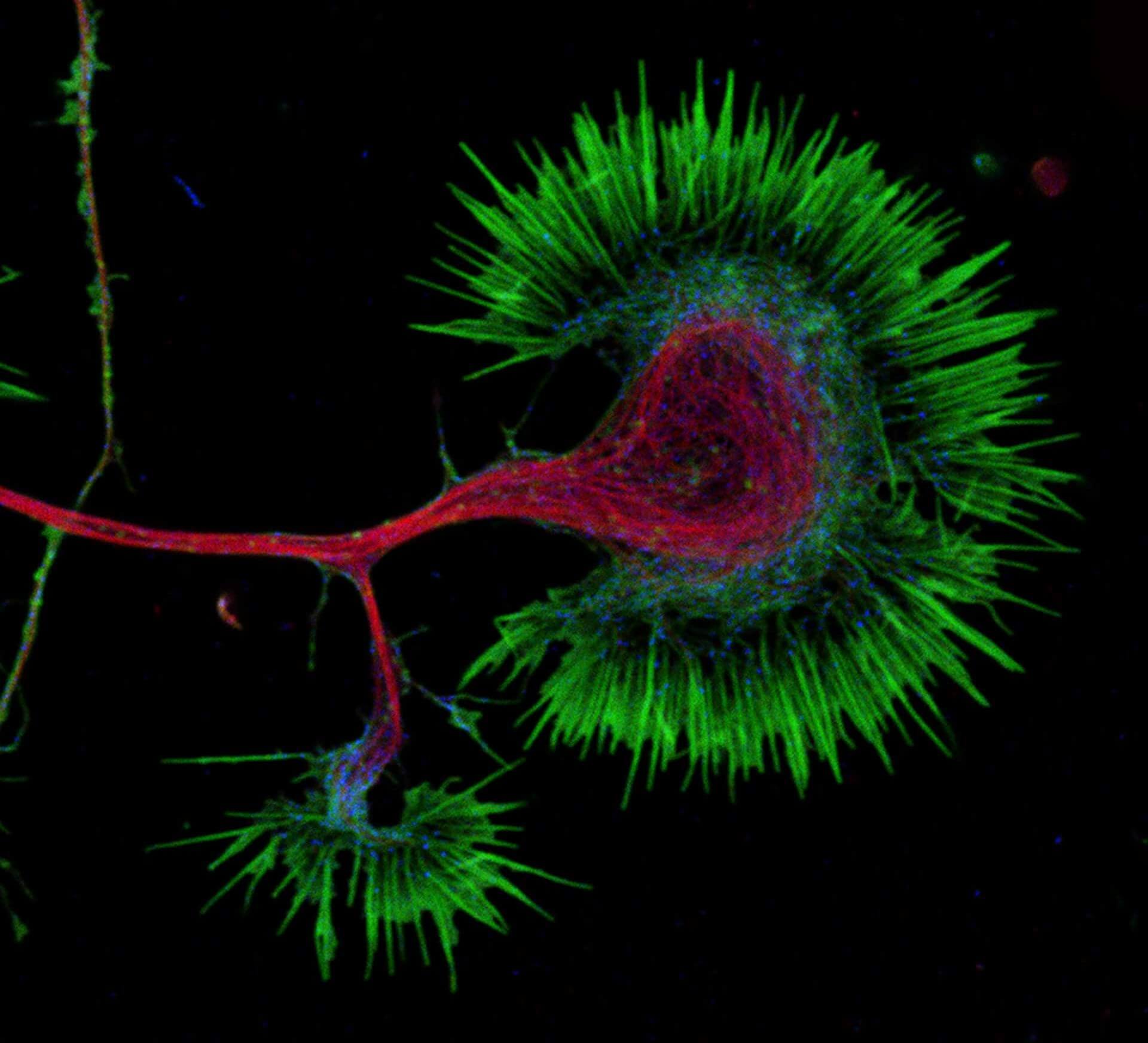
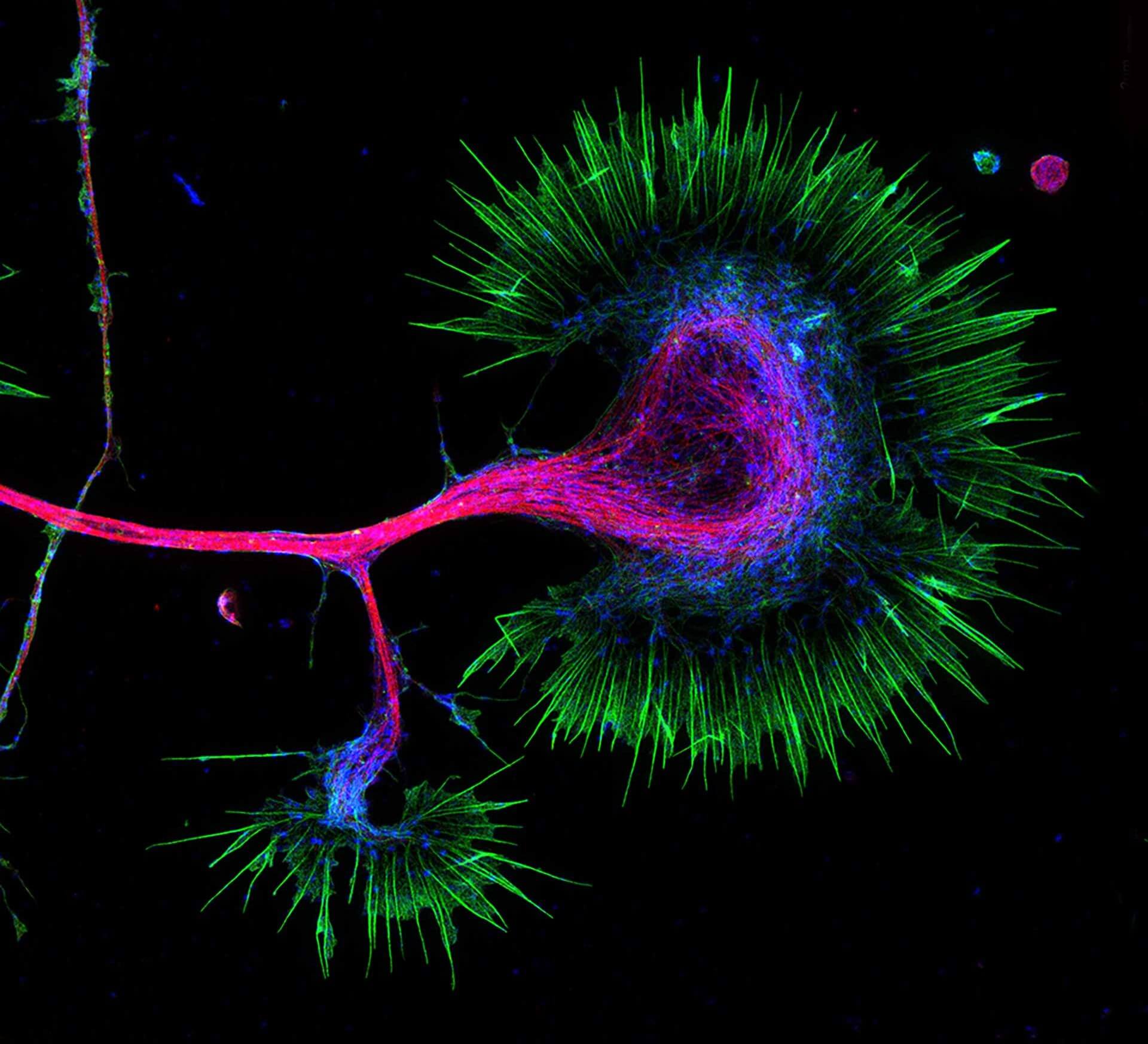
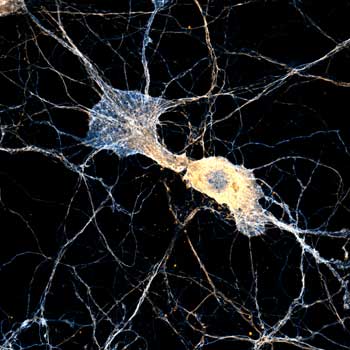
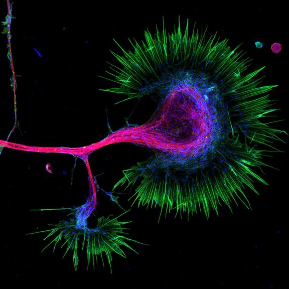
Description
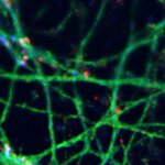
Growth cone at the tip of the axon of a primary hippocampal neuron at 1 day in vitro. Microtubules (Tuj1, abberior STAR 580, red) are bundled in the central-domain suggesting a pausing state. The molecular motor myosin IIB (confocal, Alexa488, blue) is enriched at the transition-zone, along the F-actin arcs. In the peripheral domain actin forms bundles in the filopodia (Phalloidin, abberior STAR 635, green).
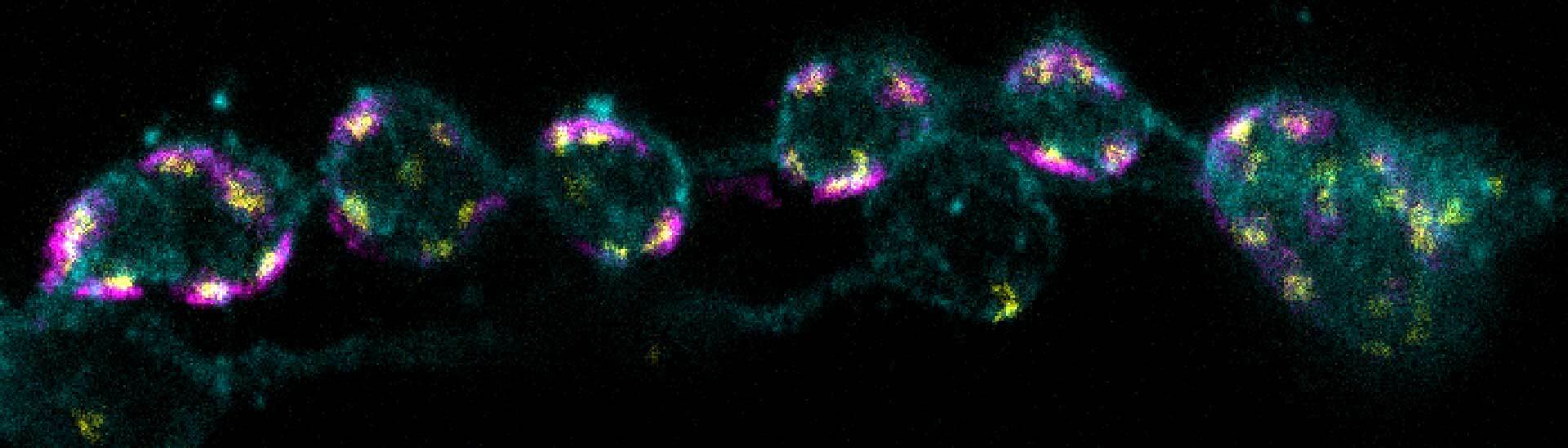
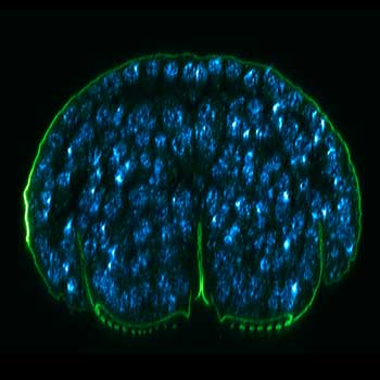
Description
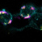
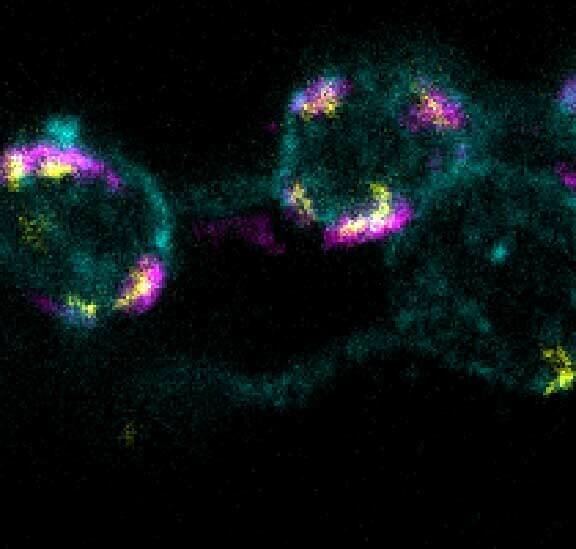
3-color STED imaging: active zones at the Drosophila larval neuromuscular junction immunostained for Bruchpilot and two other proteins.
Two superresolution channels (magenta, yellow) using a 775nm STED laser & one superresolution channel using a 595nm STED laser.
Samples by M. Lenz & M. Landgraf (University of Cambridge, UK).
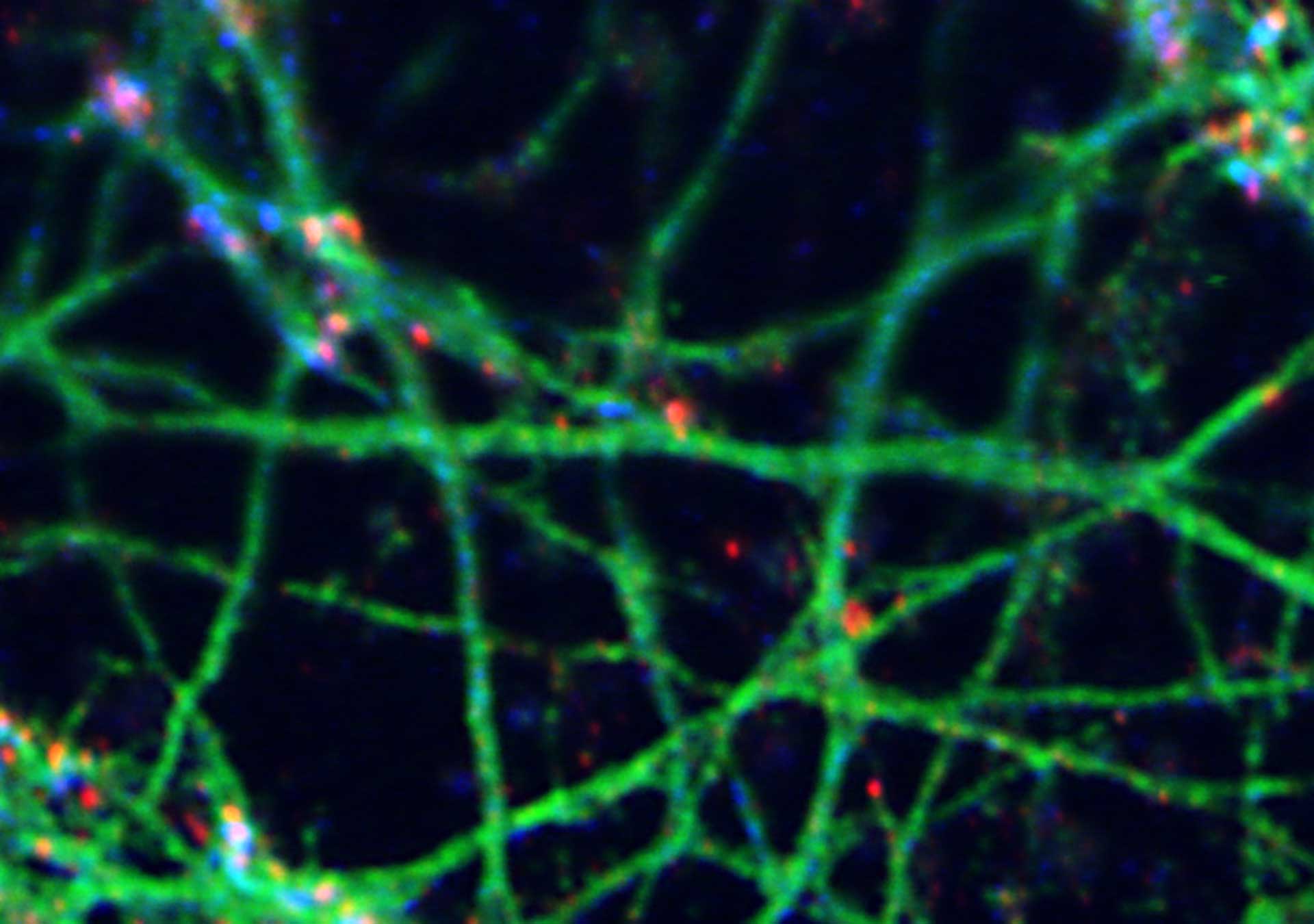
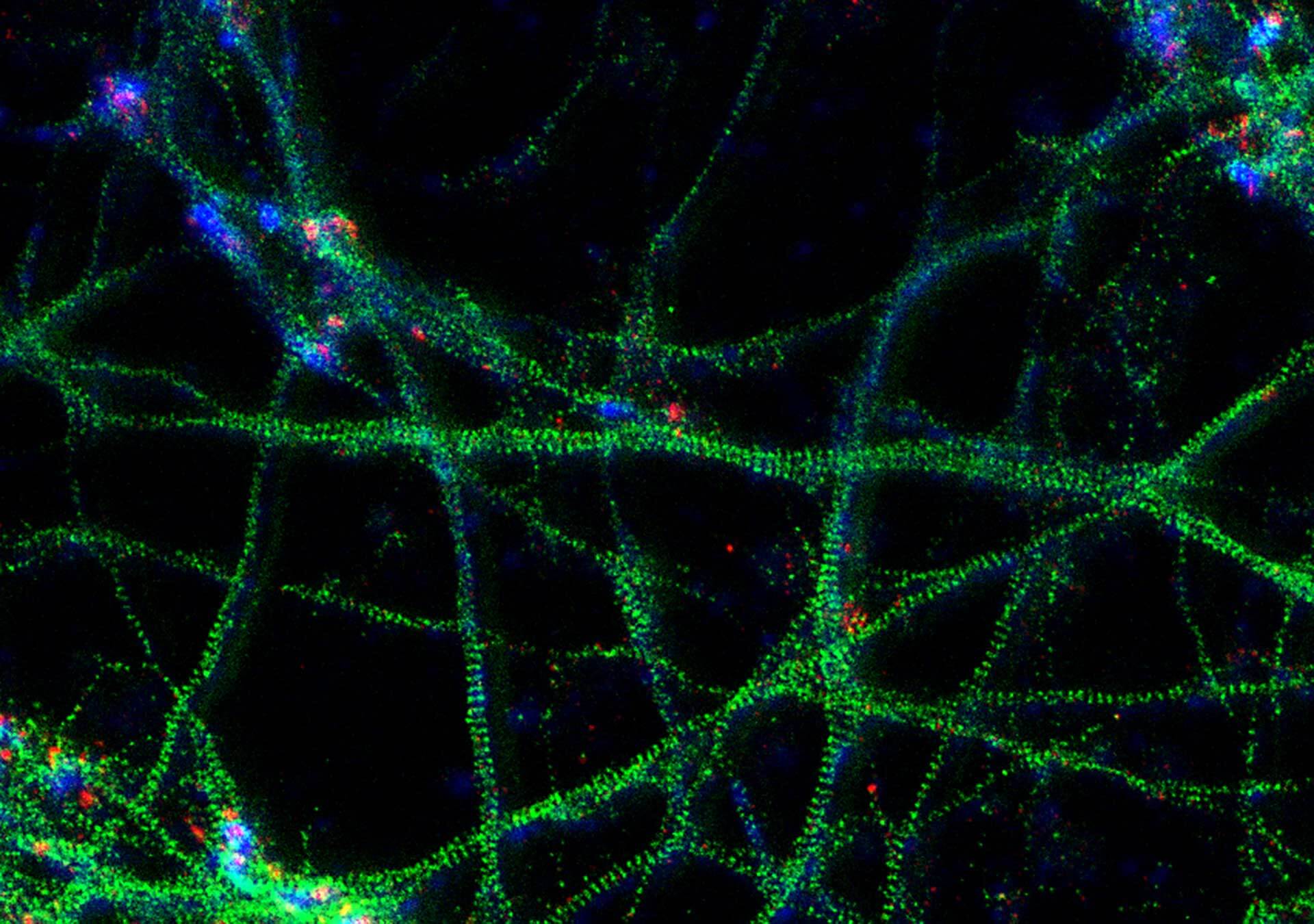
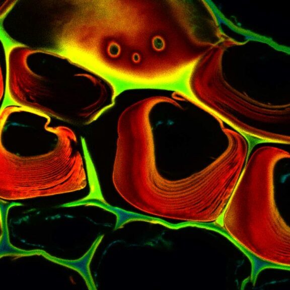
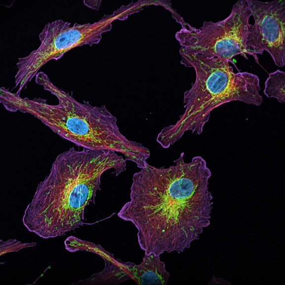
Description
3-color STED image of primary hippocampal neurons. Please note the characteristic ~190 nm beta II spectrin periodicity along distal axons (green) which is only visible in the STED image. Labelled structures: beta II spectrin (green, abberior STAR 635P), Bassoon (red, abberior STAR 580), Actin cytoskeleton (blue, phalloidin, Oregon Green 488). Imaged with abberior Expert Line with 595nm and 775nm STED laser. Sample was prepared by Elisa D’Este @ MPIBPC, Göttingen.
Modules:
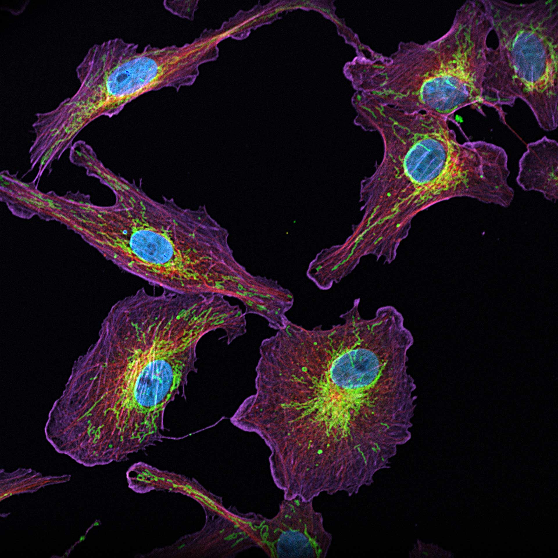
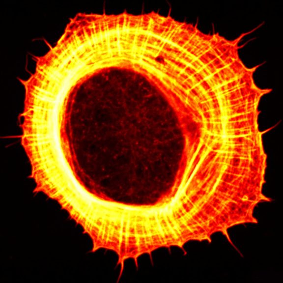
Description
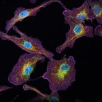
4-color confocal image of mammalian cells (DAPI, Phalloidin, Tubulin, Tom20).
Modules:


Spectral selection
with ultra-high transmission
A pair of gradient-coated filters are translated within the RAINBOW Detection in order to freely define the edges of the detection window between 400 and 800 nm. Up to four RAINBOW devices can be installed or upgraded at any time and the fluorescence can be freely distributed between every detector.
Super-sensitive
detection
Our standard detectors are avalanche photo detectors (APDs), because they have superior quantum efficiencies, up to a factor of two above hybrid detectors. This means that even when the signal levels are low, our APDs still collect plenty of photons for a meaningful image. Typical applications are superresolution STED imaging and experiments with low labeling densities designed to maintain physiological conditions. At the same time, high-signal samples are perfectly taken care of by DYNAMIC PLUS, our dead-time compensation that yields crisp images of brightly labeled samples. This way, confocal imaging is highly improved, too. Of course, you can opt between quantitative analysis of raw data or mathematical post-processing, e.g. deconvolution. Because we always give you genuine photon counts, you remain in full control of your data interpretation. Always.
Other detectors are available on request.
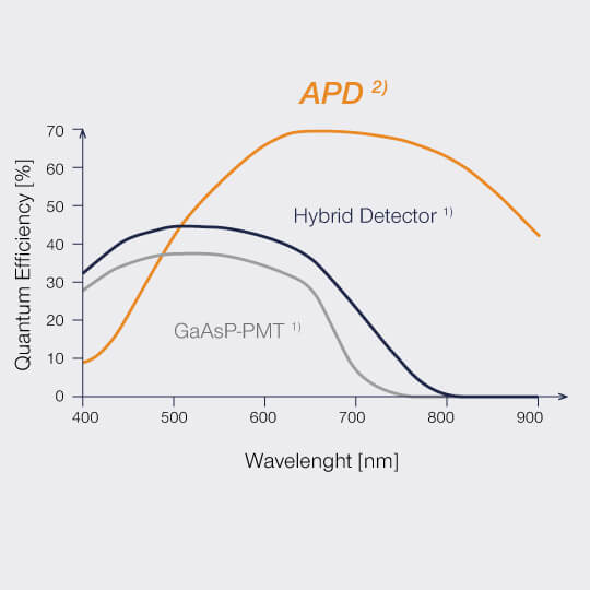
1) “Leica HyD for Confocal Imaging”, Order no.: English 1593003013 ∙ 06/2015/STO, Leica Microsystems
2) “SPCM-AQRH Single Photon Counting Module”, Rev 2018-12, Excelitas Technologies Corp.
- Gap-free spectral detection
- Choose any wavelength band between 400 and 800 nm
- APDs for the highest sensitivity (up to 65% quantum efficiency)
- Free selection of excitation and STED wavelength combinations
Because every photon counts

Why do we usually recommend APDs in our microscopes and why aren’t we worried about the supposedly lower dynamic range?
Having too many photons is never a problem. Therefore, detectors with the highest quantum efficiency are always the best choice, such as in a MATRIX array.