


Everything
Not rocket science, but simple protocols that make the daily life of a fluorescence microscopist much easier.
about labeling
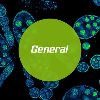
General Information
abberior offers a variety of excellent fluorescent dyes with properties optimized for the labeling of biomolecules, for spectroscopic studies and for optical microscopy, particularly super-resolution microscopy and optical nanoscopy.
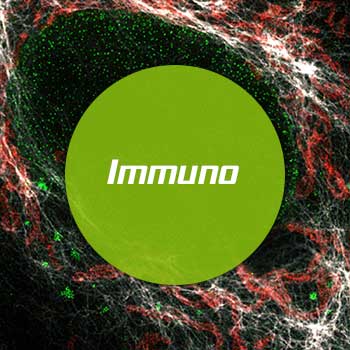
Immunolabeling protocol
Our abberior fluorescent secondary antibodies are relevant to techniques that rely on the use of fluorophore-conjugated antibodies such as flow cytometry, ELISA, western blot and immunohistochemistry.
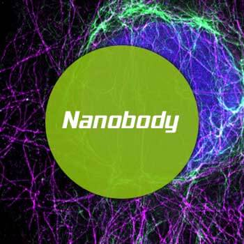
Nanobody labeling protocol
The combination of super bright and highly photostable abberior dyes and polyclonal nanobodies produced in alpacas offer a fantastic solution for high resolution imaging experiments where small labels, good penetration and outstanding specificity are necessary.
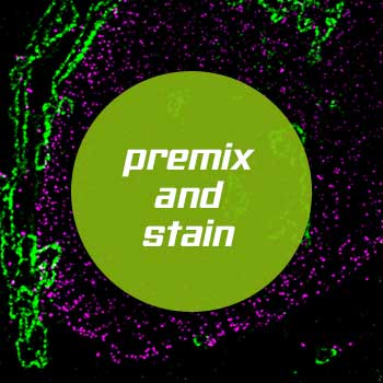
Premix & stain protocol
For very fast and easy labeling, premix a primary antibody with our abberior nanobody-conjugates and stain your biological sample in just a few steps.
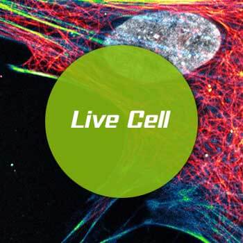
Live cell labeling protocol
Our abberior LIVE probes can be readily used to specifically label microtubule filaments, actin network, or double stranded DNA in living cells or can be used for in vitro studies.
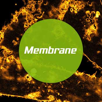
Membrane labeling protocol
Our abberior STAR membrane probes can be readily used to specifically label the outer plasma membrane of living cells. The natural behavior of the membrane and fluidity will maintain.
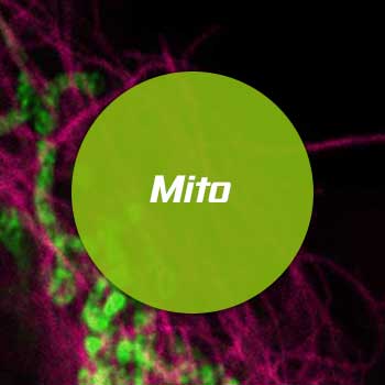
Mitochondrium labeling protocol
Our abberior LIVE ORANGE mito probe can be readily used to specifically label the christae in mitochondria of living cells. This bright and photostable probe allows the observation of natural behavior of substructures inside mitochondria with super resolution STED microscopy.
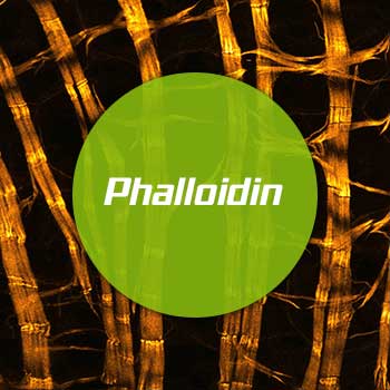
Phalloidin labeling protocol
Fluorescent phalloidin conjugates can be used to stain actin filaments (F-actin) in fixed specimens, making it an essential tool to visualize the actin skeleton of cultured cells, tissue, whole organs, or plants.

NHS ester protocol
Dyes with an N-hydroxysuccinimidyl (NHS) ester are amino-reactive derivatives and are most commonly used to label proteins such as antibodies. This reactive group helps to form a chemically stable bond between the dye and the protein (antibody).
HaloX labeling protocol
Your HaloTag® fusion proteins within living cells, on cell surfaces and on fixed cells can be reversibly colored with the abberior LIVE HaloX® labels. In a PAINT-like process imaging of the sample in an extended periods of time is easily possible, as bleaching are no longer a decisive factor.
SNAP labeling protocol
Your SNAP-tag® fusion proteins within living cells, on cell surfaces and on fixed cells can be colored with the abberior LIVE SNAP labels. These labels interact with a high affinity with the SNAP-tag® protein forming a covalent bound between label and protein.

Degree of labeling (DOL)
The degree of labeling (DOL) is the average number of dye molecules coupled to a protein molecule (e.g. an antibody). The DOL can be determined from the absorption spectrum of the labeled antibody.